''
''
A normal joint is covered in cartilage, a covering whose remarkable qualities of suppleness, elasticity and cushioning allow smooth, snag-free and, of course, pain-free movements for many, many years. Whether we are talking about mechanical parts, such as aircraft or cars, or prostheses, despite the immense progress made over the last 20 years, no technique is currently capable of reproducing a material that is both as flexible and as resistant to wear as cartilage.
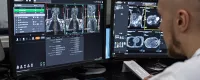
Osteoarthritis can be linked to the wear and tear of cartilage over time, with no defined cause. Example of osteoarthritis of the shoulder: note the disappearance of the space between the humeral head and the scapula, reflecting the disappearance of articular cartilage.
Pain is linked to two phenomena within the joint:
Bone-to-bone friction.
An inflammatory phenomenon:
The body reacts to the small debris worn from the cartilage by sending out inflammatory cells and producing more lubricating fluid, the joint fluid.
These two phenomena explain, on the one hand, the pain caused by prolonged use of the joint (bone-to-bone friction) and, on the other, the sometimes acute pain, with an effusion of joint fluid, the classic "synovial effusion". The effusion is therefore another sign of osteoarthritis.
It is often less visible than in the knee, for example, because the shoulder is a joint far from the skin, unlike the knee. Cracking or locking of the joints is fairly common in the shoulder. Reduced range of movement and loss of strength can be linked directly to osteoarthritis, but also to pain or a rotator cuff tear.
In a certain number of cases, osteoarthritis causes little pain in the daily activities of life and can be treated with simple medication:
Painkillers :
Paracetamol, dextropropoxyphene-paracetamol
Anti-inflammatory drugs :
Non-steroidal anti-inflammatory drugs, which must be prescribed in consultation with a doctor.
Re-education is often useful, sometimes in a swimming pool, combined with ultrasound and regular application of ice. Its usefulness and frequency must be determined by the doctor according to each particular situation.
If these initial treatments fail, one or more intra-articular injections of non-steroidal anti-inflammatory drugs may be suggested.
In practice, 3 injections a year spaced a few weeks apart seem sufficient. Only if this second stage of treatment fails should surgery be considered.
This involves inserting a camera into the shoulder through a small incision less than 1 cm long and washing the shoulder of the bone and cartilage debris that promotes inflammation and erosion of the remaining healthy cartilage. This cleaning is carried out through a second anterior incision of the same size. The operation is short (30 minutes) and can be performed on an outpatient basis.
Complications are rare, if not exceptional. The shoulder is not immobilised and can be used, with caution, as soon as the local anaesthetic is removed. The results are often good for a few months, but do not last because the wear and tear is always present, as is bone-to-bone friction. A recurrence of pain is therefore the rule after a few months. It is, however, a good waiting solution before a more serious operation.
Total or partial shoulder prostheses have made considerable progress over the last 10 years. They are much less common than hip replacements. Nevertheless, the results of modern prostheses are now excellent.
More than 90% of total shoulder prostheses are still in place more than 15 years after the operation, without pain.
Complications are also rare, but must be prevented by pre- and per-operative precautions and careful post-operative monitoring:
Rare (0.5%), infection is always a possibility, especially if infectious sites elsewhere in the body are overlooked (panariasis, dental abscess, sinusitis, urinary tract infection, infected wound, etc.) or if the body is weakened by another disease (diabetes, immunodepression, chemotherapy, etc.). If it occurs, the prosthesis often has to be removed, treated with antibiotics for several weeks, and a new prosthesis fitted once the infection has healed.
This is a stiffness of the shoulder which can also occur and has no particular specificity in a total shoulder prosthesis. FRACTURE UNDER THE PROSTHESIS This is always possible, especially in the event of a fall.
Rare (1%)
Can occur either as a result of inappropriate exertion too early, before postoperative healing, or over time if the rotator cuff is not in good condition when the prosthesis is fitted.
This is the main cause of prosthesis replacement in the long term, but remains rare (only 6% of wear requiring prosthesis replacement after 15 years of use).
Certain causes of osteoarthritis, such as bone necrosis, allow partial prostheses to be fitted, as only the humeral side of the joint is worn.
However, most cases of shoulder osteoarthritis lead to wear and tear of both the humerus and the scapula and require the fitting of total prostheses, which replace both sides of the joint.
Certain types of osteoarthritis, where the rotator cuff has been ruptured, require a special type of prosthesis, known as an "inverted" prosthesis.
Surgery for a total shoulder prosthesis requires 4 to 7 days' hospitalisation, depending on the particular case. The shoulder must then be immobilised, with the arms at your side, for three to four weeks, while the muscles heal. During this time, small pendulum movements and gentle mobilisation are carried out exclusively under the supervision of the physiotherapist. Re-education is then intensified until the shoulder regains function compatible with daily life. A stay in a rehabilitation centre is sometimes useful, especially for people who live alone or who do not have a physiotherapist close to home. Total shoulder prostheses do not give you a "new shoulder", but they do allow you to regain virtually pain-free mobility of the shoulder.
Your condition requires your shoulder to be stabilised. Nowadays, this is a well-established procedure that is increasingly practised because of its reliable results.
The shoulder is a special joint because it is not very congruent (interlocked), which explains the range of movement it is capable of (a different mobility/stability compromise to that of the hip, for example). As a result, it is vulnerable and therefore susceptible to dislocation, tearing the few existing restraints in the process: the pad, capsule, ligaments and sometimes the rotator cuff muscles. All these injuries heal poorly or not at all, which explains the recurrence of dislocations, residual apprehension or pain in certain movements.
This instability often occurs after a trauma during a countered arm movement. In rare cases, it occurs without prior trauma or after minor trauma. It is in fact an abnormal laxity of the joint which may be constitutional (abnormality of the ligament fibres) or acquired (throwing sports).
Simple elbow-to-body immobilisation is used for the first episode of dislocation for an average of 3 weeks. The principle is to relieve the patient and to hope that the anterior detachment will heal in order to prevent recurrence. Subsequently, in the event of recurrence, immobilisation is only used for pain relief for a few days (as the detachment pocket is no longer able to heal).
Afterwards, the first option is a rehabilitation programme designed to strengthen the shoulder's dynamic stabilisers (rotator cuff, subscapular muscle) while preserving the damaged static stabilisers.
However, it should be borne in mind that re-education of this poorly interlocked joint is only effective to a limited extent (depending on the type of instability, the extent of joint damage and other factors such as age, for example).
If necessary, the orthopaedic surgeon can choose from various procedures depending on the specific case to be treated, in order to achieve the best mobility-stability compromise legitimately requested by the patient.
There are 2 main types of operation for the treatment of so-called "traumatic" instability: The Bankart procedure consists of reinserting the rim and ligaments on the anterior edge of the glenoid using screw anchors, thus eliminating the anterior detachment pocket. The procedure can now be performed arthroscopically, which reduces scarring and improves functional recovery.
This type of delicate technique has been in full expansion over the last 10 years and its indications are now well defined. The Latarjet procedure consists of interposing a double obstacle in front of the potential passage of the humeral head forward: a bony stop taken at the expense of the coracoid (screwed onto the glenoid) to which the coraco-biceps remains attached, acting as a muscular stay in the dislocation position.
This type of operation requires the same type of opening (around 5cm) as the Bankart operation when performed "open". For the treatment of so-called "atraumatic" instability, the treatment is as non-surgical as possible. However, when rehabilitation proves ineffective, a procedure designed to reduce the volume of the joint (capsular plasty) may be proposed. This procedure can be performed "open" or, for some years now, under arthroscopy.
The aim is, of course, to obtain a normal joint, i.e. one that is stable, with normal, pain-free movement. Unfortunately, the shoulder is a fairly sensitive joint and the "right compromise" is not always easy to find.
Concerning stability: If we consider only recurrences, the figures vary between 5 and 10% for Bankart or Latarjet operations. If we are more demanding, taking into account minor instability accidents and residual apprehensions, 80% of shoulders are perfectly stable.
Function: Resumption of sport depends on the type of sport practised (between 3 and 6 months). The persistence of apprehension and/or limitation of external rotation usually explains the poorer results. Concerning pain: On average, regardless of which of the two techniques is used, 10% of patients who undergo surgery experience pain due to climatic conditions, fatigue or forced movements.
Short-term morbidity (complications) is fairly low (apart from the risks inherent in any hospitalisation, any stay in the operating theatre, any anaesthesia): haematomas are rare, as are technical problems during the operation, and infectious and neurological complications.
Medium-term morbidity is represented by complications such as pseudarthrosis (lack of consolidation), lysis (atrophy) or fracture of the graft in Latarjet operations. The impact of these complications on the result appears to be fairly moderate. We should also mention the possible mobilisation of implants (Latarjet screws, Bankart anchors).
Long-term morbidity, in the form of osteoarthritis of the joint, is certain but rare. Moreover, this osteoarthritis is rarely advanced and most of the time has little clinical impact.
First precaution: Maintain support with a flexible waistcoat for the entire duration indicated (the skin heals much more quickly than the muscles or bone, so do not feel "healed" too quickly).
Second precaution: Careful recovery of the various areas of mobility in rehabilitation (especially external rotation).
Third precaution: Return to sports even more cautiously, based on apprehension and avoiding sports where there is a risk of violent counter-armour (handball, volleyball).
Fourth precaution: Clinical and radiological check-ups with your surgeon for one to two years. Over the last 15 years, many authors have taken an interest in the management of shoulder instability (numerous theoretical advances, dismemberment of anatomical variants, technical improvements, arrival of arthroscopy).
Nevertheless :
The shoulder is the joint between the scapula and the humerus. The tendons that connect the muscles to the bone make up the rotator cuff. They fit around the head of the humerus and are involved in raising the arm and rotating the shoulder. The acromion is a part of the shoulder blade that forms an arch over the joint.
When the arm is raised, the tendons come into contact and rub against a projection on the acromion, which sometimes forms a real beak of bone. This repeated contact can lead to tendon rupture. The rupture of one or more tendons is manifested by pain and difficulty in raising the arm. The rupture progressively widens.
The aim of cuff repair is to relieve pain and restore mobility and strength to the shoulder. It also helps to prevent the progressive deterioration of the joint.
It consists of anchoring the ruptured tendon in its natural position around the head of the humerus. One or more anchors are screwed to the humerus. Wires mounted on these anchors are passed through the tendon and knotted together to apply the tendon to the bone.
Finally, an acromioplasty, i.e. a resection of the bony protrusion of the acromion, is performed to give more space and prevent excessive contact with the tendon.
This procedure is performed under arthroscopy, i.e. without opening the shoulder joint. Several small incisions of 5mm each are made around the shoulder. An arthroscope, i.e. a small camera, is inserted through one of them to visualise the entire joint and in particular the tendon rupture.
Small instruments are inserted through the other incisions to perform the surgery. The operation takes an average of 1 hour. It requires hospitalisation for 2 to 3 days.
Post-operative inflammatory reactions may cause significant pain and slow down rehabilitation. These exacerbated reactions sometimes correspond to algodystrophy.
Although rare, this complication takes a very long time to heal. Infection of the joint is exceptional, as the surgical procedure is performed under arthroscopy. This complication requires the shoulder to be washed and treated with antibiotics for varying lengths of time, possibly followed by a repeat operation.
Persistent joint pain or failure of the tendons to heal in the bone are also possibilities to be considered, although they are inherent in the initial pathology.
Social Services: 01-48-44-28-17
Telephone helpline 8.30am to 12.30pm and 1.30pm to 5pm
(not available on Thursday mornings, closes at 4pm on Fridays)
Walk-in service: 9.00am to 12.00pm and 1.30pm to 5.00pm
(not available on Thursday mornings, Fridays closed at 4pm)
Monday to Friday
An elbow to body splint is used to immobilise and protect your shoulder for 6 weeks. During this period, a passive re-education programme will be put in place.
After the 6th week post-op, you can permanently remove your splint and begin active mobilisation of the shoulder. The aim of the work with your physiotherapist will be to recover active mobility and muscle strength in the shoulder.
It is often necessary to wait until the 3rd month before driving again. You can usually return to work between the 3rd and 6th month, depending on your profession. Healing of the tendon to the bone is not achieved in all cases; it occurs in 60 to 80% of cases.
If the tendon does not heal in the bone, the result in terms of pain is not necessarily altered, but the recovery of strength in the shoulder is not complete. The results of this technique are nevertheless very encouraging, with pain relief and improved shoulder function in over 90% of cases.
''

To make an appointment online, it's quick and easy
click on the link below and let us guide you.

''

''
Tél : 01 43 62 22 22
41-49 Avenue du Maréchal Juin, 93260 Les Lilas